Category: Revolve

  • ASCB 2015 Reveals the Next Generation of Microscopy

    One of the best aspects of flying is being apart from the world. Not only terra firma, but the troubles of life, questions of the future, and concerns of the present. Flight requires attention to instruments while in operation, but long cross country flights leave the pilot with time to sit and watch the world unfold beneath as time passes.

    IMG_6313
    Departing from San Diego @ ASCB

    Night flight brings an even more isolated perspective, where after a few minutes alone, you watch the gauges, view the small points of light from far off cities, and feel suspended in the air, utterly alone in the earth.

    IMG_6381
    Night Flight home in the cessna.

    So last night, flying home with the sun setting behind me, I had a chance to consider the past week of ASCB, and what it meant to me.

    Trade shows are hard. They are hard for the attendees and the exhibitioners. Attendees sit through lengthy talks, then browse the show floor and get bombarded by marketing, then have dinners with colleagues into the night. Exhibitors aren’t that different. Business meetings in the early morning, standing at the show floor booth all day, and then meetings and dinners with colleagues and contacts into the night. By the last day, everyone is happy to rest on the trip home!

    Yet somehow in this internet era, there’s nothing like meeting face to face, to share life, to discuss new projects, and meet new people. This year was my 14th year of attending trade shows as a member of the microscopy community. In that time things have changed in many ways, and are exactly the same in others. I hunted through the Blanco archives and found a few shots from my very first trade show. What an impression it made on me! This was from Neuroscience 2001, with my good friend Will Casavan. I was there working for Technical Instrument Company at the time, and Will was with Media Cybernetics. DSC00008

    Little did I know that it would be 14 years later, that Will would link me up with Echo labs, and a new and exciting chapter in my life would begin.

    I bring up the past for two reasons. First, because I enjoy looking back to see how far this industry has come. 14 years ago the hottest product on the market was the Nikon TE-2000, and it sported 2, yes 2!, cameras. One for fluorescence, and one for brightfield. The inverted microscope with cameras, computer, capture cards (yes, the cameras required a card, check out that data cable!), monitor, supporting electronic boxes and cables, took up a large bench worth of space, and ran about $65,000.

    DSC00005
    Noelle looking at a TE-2000 microscope equipped with fluorescence and brightfield cameras

    Some might argue that this industry hasn’t evolved quickly enough, but consider the following. Almost every function performed on the microscope above can also be performed using the ECHO Revolve. Of course, while at the booth I was able to airdrop images like this one straight to my phone, allowing me to easily post them on the blog!

    Image_0159_zoverlay
    Three channel fluorescence captured and airdropped to my iPhone in about 30 seconds using the Revolve microscope

    To me, this is a major step forward in research technology. I think the attendees who visited the ECHO booth would agree, as while working the booth I don’t think there was a single minute without at least 1 person looking at the scopes. In 2 of the 3 days I worked the booth, we couldn’t even break for lunch!

    IMG_6262
    Yes, I was there, yes this is a selfie, don’t judge me 🙂

    While writing this, Eugene Cho, CEO of Echo labs, sent me a great timelapse of the show. I think this is an excellent video, as it shows the additional hard work required for exhibitors, to set up and tear down these displays. ECHO hand made the booth for the show, and the personal touch really made the booth stand out from the crowd in my opinion!

    Of course, there are many other advances taking place in the industry! I got to speak with Silvia Foppiano of Oko Lab, and was amazed how far incubation technology has come! In 2001, the best incubators I remember were homemade ones. Today, you can buy a stage top incubator, with magnetized connectors, micro-scale thermistor temp probes, humidity and co2 control, for prices around 1/3 of units in the past. As a machining junkie, I was very impressed with the finish of these units. If you are looking for an incubation solution you should definitely check them out.

    IMG_6290
    Magnetized hold downs keep the chambered slide in place when being used with immersion oil
    IMG_6298
    This little controller provides control and capture of all sensor data in the system.
    IMG_6292
    Chamber has perfusion ports for dynamic experiments

    Zeiss has an awesome Oculus Rift-equipped demonstration of interactive sample observation. I couldn’t believe it when I walked by, there was the rift, and no-one was sitting in the demo chair! Boom 🙂 I had to jump on that chance. As a tech nerd and gaming junkie, I’ve been relishing the chance to test these, and it was a super cool experience! At first it’s disjointed to turn and see the image field move with the accelerometer inputs on the rift. Navigation was performed using an XBOX 360 controller.
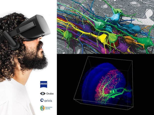
    Zeiss ad for the Oculus Rift Cellular Visualization demo

    In only a few minutes I found my senses immersed in the environment, and was soon able to fly around with ease without even thinking about the control inputs required to do so. It really was like being in another environment. Excellent demonstration by Zeiss, and I’m happy to see that these types of technology are being adapted for use in biology.

    Sutter Instrument Company had the new IPA Integrated Patch Amplifier system on display at the show. This system is a huge leap forward for cell physiologists. With a single system, you get a matched probe, amplifier, and digital capture device, all optimized for low noise and maximum sensitivity. Definitely a major step for whole cell analysis!
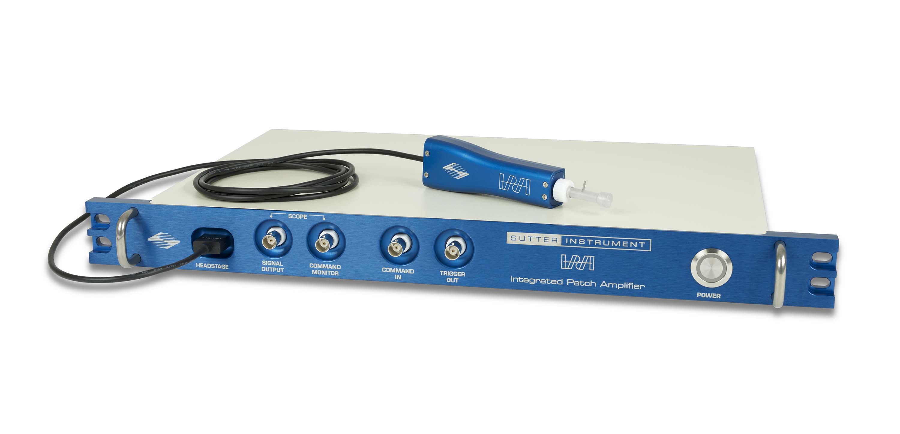
    Sutters new IPA cell patch system

    Open-Imaging had a booth as well, demonstrating the new Micro-Manager 2.0 open source microscopy platform. IMG_6221

    I’ve seen a lot of new (read unstable) software at shows like this, and was surprised at how smoothly things worked. It’s obvious that Chris and Mark have put lots of time into this version, to make sure there’s minimal chaos factor when end users upgrade. I was also able to spend some time talking with Mark, as we flew to the show together this year! I’m excited for the future of Open Imaging, and am looking forward to working with the new release!

    IMG_6200
    Mark Tsuchida takes the controls of a Mooney 201. We were speeding to the show at about 225MPH when I took this. Mark flew great!

    I want to give a special thanks to Sutter and Open Imaging, who both allowed me to present my Triggerscope at the show. I hope you got a chance to see it!

    My second observation is more a felt sense than a metric. Maybe it was the new product advancements, or my age, or who I spent tim with, but I felt a shift in generational control of the industry. During my early years in microscopy, there were a group of highly intelligent, capable and dedicated people who, in one form or another, directed the outcome of products and services in this industry. But now, a decade and a half has gone by, and I’ve realized that a new group of people are influencing that direction. While I may never get to place my personal mark on the industry, I know that I get to work near those who do.  To the people who led us here, I’d say thanks for working so hard to bring this industry such incredible capability. To those who now have the burden of keeping things new, I say bear this responsibly, not lightly. Our industry should be the hallmark of the highest ethical standards, have the strictest dedication to accuracy, and consistency. Our marketing should keep people captivated while only promising what we can deliver. We should price products fairly, and support them well. I hope we are up to the challenge, because it’s in our shoulders now.

    Yet…..when I did get a chance to walk the show floor this year, I took heart. I can see that the future is bright 🙂

    Austin

    IMG_6282
    Sunset after the show, from the top of the SD convention center


  • Echo Laboratories launches the Revolve Microscope

    Over 450 years have seen the compound microscope evolve into an incredible instrument. From simple contrast viewing, we’ve moved to super resolution systems capable of sub-diffraction accuracy. But for all this advancement, we’ve been stuck with the architecture of the microscope platform. Over 70% of labs end up buying both inverted and upright microscopes….until today!

    5 Revolve Rotation_2I’m happy to introduce the Revolve Hybrid Microscope! A scope that combines both inverted and upright observation into one instrument. I’ve worked in this industry for over a decade, and this is by far the biggest evolution in the idea of a scope I’ve ever seen! I’m so excited to share this with the research community, and I hope you’ll enjoy learning more about it!

    The Concept

    We all know that uprights and inverts use similar objectives, illuminators, position systems and cameras. Why duplicate all of these expensive components? Can’t we find a way to merge these two systems into one unified instrument? Echo Labs has done just that, with the Revolve.

    RevolveOp

    As you can see above, this a completely new way of approaching what a microscope should be! The revolve is two microscopes in one. It provides a fully capable inverted research instrument, AND a fully capable upright microscope, into one device. So the revolve is 2x the microscope at 1x the price, size and maintenance. While the revolve brings so much more capability, it’s also less, in all the ways less can be good. You’ll note the lack of a tower computer. It’s gone! The integrated iPad drives everything inside the scope. No more rat’s nest of wires on your lab bench. Of course, with the iPad, training isn’t required for new users. Both my boys (6 & 7yrs old) got a chance to use the fluorescent side of the app, and were capturing multichannel fluorescence with a few seconds. Using the touchscreen is simple and an easy extension to the other controls on the scope, so the keyboard and mouse won’t be missed. You may also note a lack of wires. In normal marketing pictures microscope manufactures won’t connect all the boxes, so things “look clean”, but the revolve IS clean. Only 1 power wire is required for operation, and all of the other brightfield, and fluorescent components are placed inside the body of the scope! To see these functions live, Echo Labs has produced a great website which includes demonstration videos, at www.echo-labs.com 

    The Revolve Microscope

    Revolve

    Let’s get right to the specs. This is a compound, infinity-path microscope. Current glass selection includes the entire line of Olympus objectives, with everything from long working distance phase to high NA oil immersion lenses. Both a high NA and long working distance transmitted light condenser are available to support phase and brightfield. The instrument uses the Apple iPad for control, interface, storage and display of images. The body and chassis are designed to be compact, fitting inside a fume hood or a lab bench with shelving installed. A single power supply runs all internal components, so there is only a single power connection required, and aside from that, no other cables need to be connected.

    Brightfield

    In brightfield mode the iPad camera is optically coupled to the lightpath, providing sharp, color balanced, auto exposed images. Optical coupling is set to provide the full objective field within the iPad camera FOV, with pinch+zoom available should the user want a traditional square view. On-screen controls are provided for color balance, brightness and contrast.

    IMG_9727 (1)

    Due to the unique nature of the upright/inverted combination, both high res close working condensers as well as long working distance phase condensers are available. A high accuracy locking mechanism is used to securely hold the condenser assembly in place, while still allowing for easy removal by a single lever.

    Fluorescence

    In fluorescent mode the Revolve uses a high sensitivity quantitative monochrome camera, which is wirelessly connected to the iPad display, providing a no-latency view when scanning a fluorescent plate or slide. High power discrete LED’s provide fluorescence excitation. Fluorescent wavelengths are specified by “light cube”, with a cube and filter block set provided for popular fluorescent excitation/emission spectra.

    IMG_9761

    The user interface for fluorescence includes enough control to be useful, but avoids the excessive detail usually found on older imaging programs. You won’t have to worry about parallel shift voltage settings, or pre/post sensor clearing options just to snap an image! On-image display scaling, a simple high/low gain selection, and attenuation control are easily controlled. The physical knob on the right side of the microscope controls the current fluorescent channel in parallel with the app, providing a bidirectional link for the user’s preferred mode of operation.

    Mechanical

    The body includes a USB hub which can accept common memory sticks, allowing for quick USB transfer of images obtained by the iPad app. The engineers spent an extensive amount of time working on the question of “what do we do with the images on the iPad?”. This is an important question for every microscope, yet it never seems to be completely addressed. How many times have you realized images you captured simply were lost, because someone deleted them from your scope’s hard drive! With the Revolve you can save your images almost anywhere: USB, AirDrop, DropBox, with more cloud options coming soon. Or, just take your iPad….and walk away!

    Revolve side

    The focus mechanism is duplicated for easy access in either upright or inverted configuration, providing a common coarse/fine knob adjustment. X/Y adjustment is provided by dual knob control placed close to the user’s hand. The stage is locked into the microscope with a secure, but easily removable cam lock and alignment pins. This makes removal of the stage easy for revolving the scope, but keeps the stage securely in place when in use. Check out this video for an example of each of the above functions!

    Conclusion

    During my time working with the Echo Labs team, I’ve seen the attention to detail put into the user-focused features of this microscope. My previous posts have hopefully allowed a sense of understanding as to why I am so excited to work with this team! This company is a completely new approach to an old industry. The management team consists of veterans in the microscopy industry with a passion for high end imaging. The engineers are an amazing cadre with broad experience in the music, medical, defense, and research industries. Watching this team put the combined skill and experience into this microscope has been simply incredible. I hope you get a chance to experience using this microscope, as you’ll never look at scopes the same way again! Of course, in keeping with the idea of a new-generation company, you don’t need to talk with anyone just to check out pricing, an online and interactive quote configurator is available, which allows you to easily build the revolve for your work.

    I believe Echo labs is well on the way to completely changing the microscopy industry. The Revolve is only the first step in bringing easy to use, fair price research instruments into the lab. Welcome to the Revolution.

    -Austin Blanco


Social Media Auto Publish Powered By : XYZScripts.com